

 SkillClick
What Is the Difference Between a Cyst and a Cancerous Skin Lump?
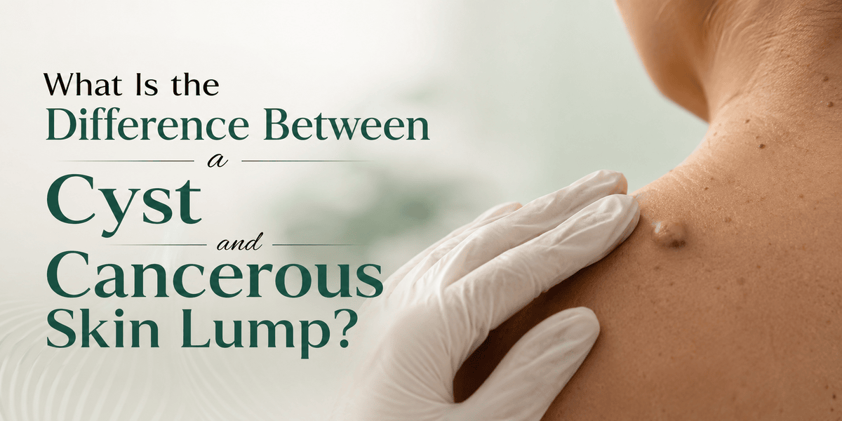
Have you recently discovered a lump beneath your skin and found yourself wondering whether it's a harmless cyst or something more serious? It's a concern that crosses many people's minds, especially when a lump appears unexpectedly or begins to change over time. While most skin lumps turn out to be benign cysts, some may require prompt medical assessment to rule out skin cancer. Knowing the differences can help you seek appropriate care without unnecessary worry.
Skin lumps are common, yet distinguishing a benign cyst from a potentially cancerous growth can feel uncertain. Cysts typically develop as closed sacs filled with keratin, fluid, or other material, whereas cancerous lumps result from abnormal cell growth that can invade surrounding tissues. Although some cysts remain harmless and never require treatment, others may become painful, infected, or cosmetically bothersome, making cyst removal surgery an appropriate option in certain cases. Dermatologists use clinical examination, medical history, and, when necessary, imaging or biopsy to distinguish between these conditions and recommend the most suitable course of action.
This article explains the practical differences between common skin cysts and cancerous skin lumps, including how they look, feel, and behave over time. It also highlights the warning signs that warrant professional assessment, outlines how dermatologists reach a diagnosis, and discusses when observation, treatment, or removal may be recommended.
Your skin often reveals small changes long before they become major concerns.
What Are Skin Cysts and How Do They Form?
Skin cysts are closed, sac-like structures that develop within the dermis or subcutaneous tissue. The most frequent types are epidermoid cysts (sometimes called sebaceous cysts) and trichilemmal cysts. They form when keratin or sebum becomes trapped beneath the skin surface, often after a hair follicle or gland opening becomes blocked. The cyst wall continues to produce material, causing the lump to enlarge slowly over months or years. Most cysts remain soft or firm, movable, and painless unless they become inflamed or infected. They are not contagious and carry no inherent tendency to become cancerous. Understanding this mechanism helps explain why cysts feel different from solid tumours and why they sometimes fluctuate in size or develop secondary infection.
What Does a Typical Skin Cyst Look and Feel Like?
Recognising the classic features of a cyst supports early self-assessment and reduces unnecessary anxiety.
These characteristics are observed consistently during clinical examinations and help distinguish cysts from other lumps.
- Shape and Surface: Usually round or dome-shaped with a smooth surface. A small central punctum (dark pore) is often visible on epidermoid cysts.
- Texture and Mobility: Soft to firm, and freely movable beneath the skin. The lump can often be shifted slightly from side to side.
- Growth Pattern: Slow enlargement over months or years. Sudden increase in size usually signals inflammation or infection rather than aggressive growth.
- Colour: Flesh-coloured or slightly yellowish. Redness appears only when the cyst becomes inflamed.
- Symptoms: Most are asymptomatic. Pain, tenderness, or discharge of thick, cheese-like material occurs if the cyst ruptures or becomes infected.
- Common Locations: Face, neck, scalp, trunk, and areas rich in sebaceous glands or hair follicles.
- Number: People may have a single cyst or several; multiple cysts can occur without indicating systemic disease.
Clinical Insight
Dermatologists frequently note that patients worry most about firmness or recent growth. In practice, a smooth, movable lump with a visible punctum is far more likely to be a cyst than a malignant tumour. However, any lump that feels fixed to deeper tissue, grows rapidly, or develops ulceration warrants careful examination to exclude less common diagnoses.
What Are the Main Types of Cancerous Skin Lumps?
Skin cancers present in several forms, each with distinct clinical patterns. Awareness of these patterns aids recognition of lesions that need urgent review.
Basal Cell Carcinoma
Basal cell carcinoma is the most common skin cancer. It often appears as a pearly, translucent nodule with visible blood vessels, or as a non-healing sore. Growth is typically slow, and the lesion may ulcerate or bleed intermittently. It rarely spreads beyond the local site but can invade nearby tissue if left untreated.
Squamous Cell Carcinoma
Squamous cell carcinoma may develop as a firm, red nodule or as a scaly, crusted patch that fails to heal. It occurs more frequently on sun-exposed skin and carries a higher risk of local invasion and, less often, distant spread than basal cell carcinoma.
Melanoma and Other Less Common Tumours
Melanoma usually arises in pigmented lesions and shows the classic ABCDE features: asymmetry, border irregularity, colour variation, diameter increase, and evolution. Other rare malignant lumps include Merkel cell carcinoma or cutaneous metastases from internal cancers; these tend to grow more rapidly and feel firmer or more fixed.
How Do Cancerous Lumps Differ in Appearance and Behaviour?
Cancerous skin lumps display several features that contrast with the typical behaviour of cysts.
Observing these differences helps prioritise which lesions require prompt medical attention.
- Growth Rate: Malignant lesions often enlarge more rapidly over weeks to months, whereas cysts grow slowly over years.
- Surface Changes: Ulceration, crusting, bleeding without trauma, or a non-healing appearance raise concern. Cysts may discharge keratin but usually do not ulcerate spontaneously.
- Fixation: Cancerous lumps can become tethered to surrounding tissue and feel less mobile. Cysts remain freely movable in most cases.
- Colour and Border: Irregular pigmentation, multiple colours, or poorly defined borders are more characteristic of melanoma or other atypical lesions.
- Symptoms: Persistent pain, itching, or spontaneous bleeding without infection is more common with malignant growths.
- Associated Skin Changes: Surrounding redness, scaling, or satellite lesions can accompany certain skin cancers.
- Response to Simple Measures: Inflamed cysts often settle with time or antibiotics; malignant lesions continue to progress.
These clinical clues guide the decision for further investigation.
Which Warning Signs Suggest a Lump Needs Urgent Assessment?
Certain features should prompt timely medical review rather than continued observation. Rapid growth over a few weeks, ulceration or spontaneous bleeding, fixation to deeper structures, and irregular pigmentation all justify prompt examination. A lump that appears in a previously normal area after the age of 40, especially on sun-damaged skin, also merits evaluation. Systemic symptoms such as unexplained weight loss or fatigue accompanying a new skin lump are uncommon but require comprehensive assessment. Even in the absence of dramatic changes, any lesion that looks markedly different from a person’s other skin growths should be checked. Early assessment allows accurate diagnosis and, when needed, prompt management.
How Do Clinicians Distinguish Cysts from Cancerous Lumps?
Dermatologists combine history, examination, and selective investigations to reach a diagnosis.
The process is systematic and tailored to the individual lesion.
- Clinical History: Onset, growth rate, previous trauma, infection episodes, and sun-exposure history provide essential context.
- Physical Examination: Assessment of size, shape, surface, mobility, colour, and surrounding skin. Dermoscopy may reveal additional surface detail.
- Imaging: Ultrasound can confirm the cystic nature of a lump by showing fluid content and a well-defined wall. Solid or irregular masses may need further imaging.
- Biopsy or Excision: When clinical features are atypical, a sample or the entire lesion is removed for histopathological examination. This step provides definitive tissue diagnosis.
- Laboratory Analysis: Microscopy identifies the cyst wall and contents or, conversely, confirms malignant cell types and guides staging if cancer is found.
- Follow-up Planning: Benign cysts may be observed or removed electively; malignant diagnoses lead to structured treatment pathways according to national guidelines.
This structured approach minimises both over-treatment of harmless lesions and delayed recognition of serious ones.
When Is Removal Recommended for a Cyst or Suspicious Lump?
Removal decisions differ according to the suspected diagnosis and the patient’s symptoms.
Elective Cyst Removal
Cysts are often removed when they cause recurrent inflammation, discharge, discomfort, or cosmetic concern. Cyst removal surgery is typically performed under local anaesthetic as a day-case procedure. Complete excision of the cyst wall reduces the chance of recurrence. Inflamed cysts may be treated with antibiotics or drainage first, with definitive excision planned once the infection settles.
Removal of Suspicious or Confirmed Malignant Lumps
Any lesion with features suggestive of skin cancer is usually excised with appropriate margins and sent for urgent laboratory analysis. Further treatment depends on the histological result and may include wider excision, topical therapies, radiotherapy, or systemic treatment according to established protocols from organisations such as NICE and the British Association of Dermatologists.
Shared Decision-Making
The clinician discusses benefits, risks, expected scarring, and recovery time so that the patient can participate in the choice. Not every cyst requires removal, and not every suspicious lump proves malignant; accurate diagnosis remains the priority.
What Are the Risks of Delaying Assessment of a Concerning Lump?
Postponing evaluation of a changing or atypical lump can allow a malignant process to progress. Early-stage skin cancers are highly treatable, with excellent outcomes when managed promptly. Delayed diagnosis of melanoma or invasive squamous cell carcinoma may necessitate more extensive surgery or additional therapies. Even for benign cysts, repeated infection can lead to scarring and more difficult subsequent removal. While most lumps remain harmless, the small proportion that are malignant benefit substantially from early detection. Public health guidance consistently emphasises timely review of new or changing skin lesions as a practical step toward better outcomes.
Skin Health Snapshot
- The large majority of skin lumps examined by dermatologists are benign.
- Early detection of melanoma is associated with markedly higher survival rates.
- Basal cell carcinoma is the most common skin cancer and rarely metastasises.
- Any lump that grows rapidly, ulcerates, or becomes fixed should be assessed promptly.
How Can People Monitor Skin Lumps in Daily Life?
Regular self-examination supports early recognition of change without causing undue alarm.
Good lighting and a mirror help inspect hard-to-see areas such as the back and scalp. Noting the approximate size, colour, and surface of existing lumps creates a personal baseline. Photography can assist in tracking subtle changes over months. Any new lump, rapid enlargement, surface breakdown, or alteration in colour or mobility should prompt professional review. Sun-protection habits reduce the long-term risk of certain skin cancers and form a useful complement to monitoring. People with a personal or family history of skin cancer or numerous atypical moles may benefit from scheduled dermatological checks in addition to self-examination.
- Monthly Checks: Examine the skin systematically, including scalp, behind ears, and soles.
- Documentation: Note or photograph lesions that are new or changing.
- Trigger Features: Rapid growth, bleeding, ulceration, or fixation warrant prompt assessment.
- Sun Protection: Daily broad-spectrum sunscreen and protective clothing lower cumulative ultraviolet damage.
- Professional Input: Seek review when uncertainty exists rather than relying solely on self-diagnosis.
At a Harley street skin clinic, clinicians often evaluate patients who present with precisely these concerns, providing clarity through examination and, when indicated, further testing.
What Practical Steps Follow a Diagnosis?
Once a diagnosis is established, the next actions depend on whether the lump is a simple cyst or a malignant lesion. For a confirmed cyst, options include observation, aspiration in selected cases, or elective excision if symptoms persist. After cyst removal surgery, standard wound care and a short period of activity modification usually suffice. For a cancerous diagnosis, the dermatologist or multidisciplinary team outlines the recommended treatment pathway, which may involve further surgery, topical agents, or other modalities according to the specific cancer type and stage. Clear written information and follow-up arrangements help patients understand timelines and expected outcomes. Regardless of diagnosis, continued skin surveillance remains important because new lesions can develop over time.
Case Study
A patient noticed a firm, slowly enlarging lump on the upper back that had been present for two years. Recent mild tenderness led to clinical assessment. Examination showed a smooth, movable mass with a central punctum. Ultrasound confirmed a cystic structure. The lesion was excised under local anaesthetic; laboratory analysis verified a benign epidermoid cyst. Recovery was straightforward, and no further treatment was required. The episode demonstrated how typical clinical features, supported by simple imaging when needed, can distinguish a common cyst from more concerning lumps and guide appropriate, limited intervention.
Comparing Skin Cysts with Cancerous Skin Lumps
| Feature | Typical Skin Cyst | Cancerous Skin Lump |
| Growth Rate | Slow (months to years) | Often faster (weeks to months) |
| Mobility | Freely movable | May become fixed to deeper tissue |
| Surface | Smooth; possible central punctum | May ulcerate, crust, or bleed |
| Consistency | Soft to firm, often fluctuant | Firmer, solid |
| Pain | Usually none unless infected | May be tender or painful without infection |
| Colour | Flesh-coloured or yellowish | Variable; irregular pigmentation possible |
| Discharge | Thick keratin if ruptured | Blood or serous fluid if ulcerated |
| Cancer Risk | Negligible | Present; depends on specific type |
| Usual Next Step | Observation or elective removal | Prompt biopsy or excision for diagnosis |
FAQs About Cysts and Skin Lumps
Can a cyst turn into cancer?
Ordinary epidermoid or trichilemmal cysts do not transform into cancer. However, a lump initially thought to be a cyst occasionally proves to be a different diagnosis once examined histologically, which is why atypical features prompt removal and laboratory analysis.
How quickly should I have a new lump checked?
Any lump that grows rapidly, ulcerates, bleeds without trauma, or feels fixed should be assessed promptly. Slow-growing, typical-appearing cysts can usually be reviewed routinely.
Is imaging always required?
No. Many cysts are diagnosed on clinical examination alone. Ultrasound is helpful when the nature of the lump is unclear or when deeper extension needs evaluation.
What does cyst removal surgery involve?
The procedure is normally performed under local anaesthetic. The cyst and its wall are excised, and the skin is closed with sutures. Most people return to normal activities within a few days, depending on the site and size.
Will the lump come back after removal?
Complete excision of a cyst wall makes recurrence uncommon. Incomplete removal or prior infection can increase the chance of return. New, unrelated cysts can still form elsewhere.
To Bring It All Together
Most skin lumps prove to be benign cysts that develop from blocked glands or follicles and grow slowly. Cancerous lumps are less common and typically display faster growth, surface breakdown, reduced mobility, or irregular pigmentation. Careful attention to these differences, combined with regular self-examination, enables people to seek review at the right time. Dermatologists use history, examination, and selective investigations to reach an accurate diagnosis and recommend observation, cyst removal surgery, or further treatment as appropriate. Early assessment of atypical features supports the best possible outcomes while avoiding unnecessary procedures for clearly benign lesions. Ongoing sun protection and skin awareness remain valuable habits for long-term skin health. This article is intended for educational purposes only and should not replace professional medical advice, diagnosis, or treatment. If you notice concerning skin changes or have questions about your health, consult a qualified healthcare professional.
- Managerial Effectiveness!
- Future and Predictions
- Motivatinal / Inspiring
- Fitness and Wellness
- Medical & Health
- Manufacturing
- Образование
- Real-Estate
- Food Industry
- Hospitality
- Online Games
- Sports
- Home Services
- Civil Engineering
- Safety and Protection
- Software Products & Services
- Fashion and Jewellery
- Artificial Intelligence
- Entrepreneurship
- Mentoring & Guidance
- Marketing
- Networking
- HR & Recruiting
- Literature
- Shopping
- Career Management & Advancement